
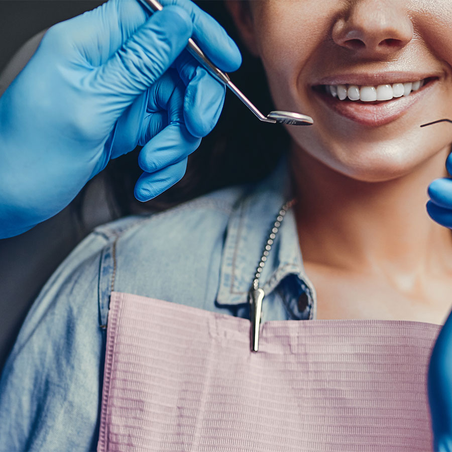
Whistler Dental is a place where the genuine care and comfort is our top priority. We pledge to provide the finest personal service for our guests who will always enjoy a safe, compassionate, trusting, and professional atmosphere.
GENERAL SERVICES
At Whistler Dental we use composite white filling materials when restoring cavities. These fillings are made of materials that harden under a special light to form a strong, long lasting and esthetic dental restoration. In the past, silver amalgam fillings were one of the only options available to restore teeth with tooth decay, but with advances in dental technology, we no longer need to rely solely on these materials. Composite white fillings have several advantages over their amalgam counterparts, including:
- Minimally invasive – we are able to preserve more of your natural tooth with these materials
- Esthetically pleasing
- Ability to bond directly to natural tooth
- Do not conduct hot and cold, potentially decreasing sensitivity
- Highly successful in moderately sized cavities
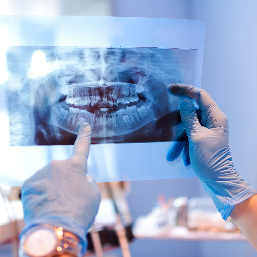
Ceph x-ray – takes pictures of the lateral or side view of the face. The complete radiographic image provides a way to view the teeth, jawbone and soft tissues that cannot be seen with the naked eye. This profile view of one’s face displays the bones and facial contours on a single film for a more accurate diagnosis. This type of x-ray is primarily used in the orthodontic planning and treatment process.
Cone Beam CT – provides detailed images of the bone and is performed to evaluate diseases of the jaw, dentition, bony structures of the face, nasal cavity and sinuses. It does not provide the full diagnostic information available with conventional CT, particularly in evaluation of soft tissue structures such as muscles, lymph nodes, glands and nerves. However, cone beam CT has the advantage of lower radiation exposure compared to conventional CT.
VELscope – VELscope is a simple, non-invasive handheld device that was developed by the BC Cancer Agency to detect oral cancer. The VELscope system: uses a safe and painless blue light to distinguish between normal and abnormal tissue. It only takes two minutes, is non-invasive and does not require any special rinses or stains.
Laser Dentistry – Laser dentistry is used for biopsies, tissue retraction around crown preparations and frenectomies. It allows for quick and non-traumatic treatment resulting in precise and sterile incisions. It is ideal for children.
Air Abrasion – this alternative to the traditional dental drill method is available for small caries to remove tooth decay, superficial stains and tooth discolorations. It is a simple and quick procedure that reduces the need for anesthesia and does not produce any heat, sound, pressure or vibrations. It is also great for children.
At Whistler Dental we are able to offer mouthguards for many situations; whether you grind your teeth in your sleep, go hard on the mountain, on the ice or in your chosen sport, or even have a beautiful new smile to protect… we have the mouthguard options to suit you. Your dentist can recommend your best fit.
Endodontics or root canals are treatments used to treat injuries or infections within the tooth. Symptoms of injured or infected teeth include; sensitivity, spontaneous or constant pain, swelling (in the gums, roof of mouth, cheek, lip or eyelid), or a bad taste in the mouth.
The procedure is performed over one or two appointments, is relatively pain-free and allows a tooth to function normally and retain its natural aesthetics, it saves teeth from being extracted. In terms of health, investment and conservative treatment, there is no substitute for retaining natural teeth. All general dentists at Whistler Dental are able to perform endodontic treatment. However, we also have Dr. Joel Fransen, Certified Endodontist who visits our office to treat complex cases. If you are experiencing any of the aforementioned symptoms, book your exam today.
At Whistler Dental we understand that not everyone enjoys visiting the dentist. This is usually based upon previous experiences. It doesn’t need to be that way; we are able to offer sedation options to take the edge off your dental experience –these sedations will not “put you under” -but, they will help you relax.
The different options are; Oral sedation, IV sedation and Nitrous (laughing gas). For more information or call us on 604 932 3677 to see if one of these options will work for you.
A dental crown is specifically designed to protect a tooth. Over the years, teeth are subject to an incredible amount of punishment. If a tooth has previously had a large filling placed in it, has be subjected to trauma or has had a piece fracture away, a dental crown may be the next best step. Dental crowns generally come in two materials- gold or porcelain. Gold crowns are durable and long-lasting, however they have the disadvantage of being yellow in colour and therefore not the best for esthetic areas of the mouth. Porcelain crowns are designed to match the colour, shape and size of the rest of your teeth and are most commonly used in dental practices today. If taken care of properly, crowns can last many years.
Getting a dental crown will usually require two appointments. At your initial visit, we will remove any broken down fillings and decay and prepare your tooth so that it can accept a crown. An impression will need to be taken of your upper and lower teeth, which are sent to a dental laboratory. In certain cases, where colour match is absolutely essential, we may ask that you visit the dental lab directly for a custom shade match of your teeth. A dental crown is then fabricated at the lab and then returned to our office. During your second appointment, we remove the temporary crown that was placed at your initial visit and replace it with a new crown, which is permanently cemented into place.
Crowns may be recommended for the following reasons;
- Repair teeth that have been broken or fractured
- Preserve teeth that are heavily decayed
- Cosmetic correction
- Protect a tooth that has had a root canal
- Preserve teeth that have previously had large fillings
Whistler Dental Clinic is proud to introduce Dr. Reza Ahmadi, Certified Specialist in Periodontics, who will be taking care of our patients’ more advanced needs regarding their gum and bone health. There is no need for our patients to travel long distances to see a specialist in the city anymore as Dr. Ahmadi would perform their periodontal assessments, diagnosis and treatments at the comfort of our own clinic. Some of these service include but are not limited to:
Dental Implants
Dental implants are great alternatives to replacing your missing natural teeth. It allows a patient to have the comforts and confidence to eat, speak, laugh and enjoy life.
Cosmetic Periodontal Surgery
Cosmetic periodontal procedures are a predictable way to cover unsightly, sensitive, or exposed root surfaces and to prevent future gum recession.
Guided Bone Regeneration (GBR)
GBR is a predictable way to regenerate bone that has been loss due to gum disease, injury or tooth extraction. This give us the opportunity to place implants that are in length and width proper location and restore the functionality and appearance of your mouth.
Crown Lengthening
Crown lengthening is a surgical procedure performed by a dentist to expose a greater amount of tooth structure for the purpose of subsequently restoring the tooth prosthetically.
Initial diagnosis is usually made by one of our qualified dentists and an in house referral is made to Dr. Ahmadi for further assessment.
Please don’t hesitate to ask your dentist at WD if you have any concerns regarding your periodontium and whether you need a referral to see the specialist.
COSMETIC SERVICES
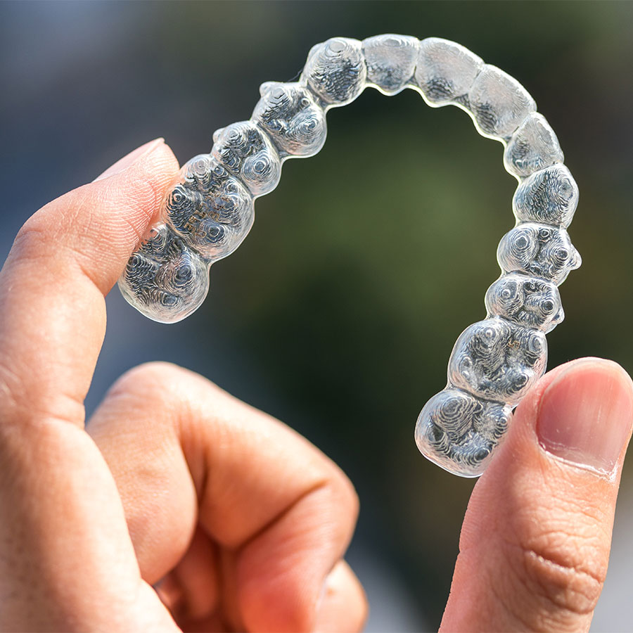
What is Invisalign?
Invisalign is an orthodontic treatment that uses clear, removable aligners to straighten your teeth without the use of traditional metal braces. The trays are comfortable, removable and almost completely invisible.
Why use Invisalign?
- Gaps or spaces between teeth
- Overbite (upper teeth bite over the lower teeth)
- Underbite (lower teeth protrude past the front teeth)
- Open bite (teeth are unable to make contact with the opposing teeth for a proper bite)
- Overly crowded teeth
- Crossbite (upper and lower jaws are misaligned)
We offer two types of whitening at Whistler Dental. In-office or take home whitening. In-office Whitening is a one-session treatment that comes with a kit to allow you to maintain your pearly whites at home.
Take-home whitening requires two short appointments in the office and allows you to whitening in the comfort and privacy of your home, on your schedule.
To see if you’re a candidate for whitening, give the office a call and we’ll get you started on your journey to a whiter and brighter smile
Dental implants are an effective, natural looking replacement for teeth that are missing as a result of gum disease, decay, failed root canal, fracture or injury. There are multiple benefits to dental implants, biologically compatible titanium is placed directly into the bone and not attached to any other teeth so they fuse safely and securely, providing long-term stability. Implants also stimulate bone growth, which prevents future facial bone loss.
How Does It Work?
Dental implants work in two stages. In the first stage, the implant is placed in the bone and left to integrate for several months. Once this phase is complete, then your implant is ready to be restored. Your dental professional will take an impression of your teeth and dental implant and have your new crown or bridge fabricated. This generally takes two weeks to complete, at which point you will receive the completed prosthetic.
Are Implants Right For Me?
If you are wondering if dental implants are the best treatment option for you, a consultation with one of our dental professionals is required. At this appointment, x-rays and a clinical exam will be performed to assess the quality and quantity of bone present at the proposed implant site. If there is inadequate bone, additional bone grafting procedures may be required before an implant can be successfully placed. We will also be happy to discuss alternatives to implants so that you are fully informed as to your treatment options.